


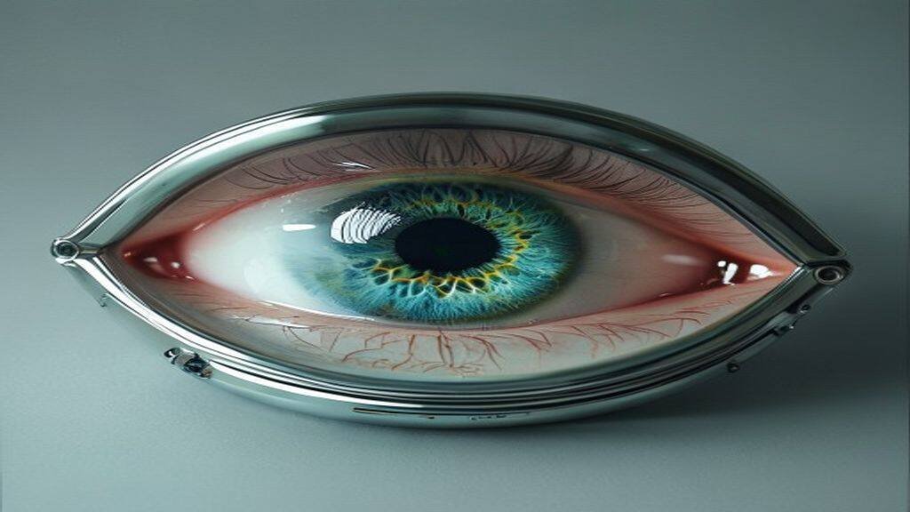
For decades, the field of ophthalmology has been tethered to a singular, frustrating limitation: the human eye is a highly complex, delicate organ that begins to degrade almost immediately upon the cessation of blood flow. While organ donation has saved countless lives, the prospect of an “eye transplant” has remained firmly in the realm of science fiction. However, a groundbreaking development emerging from the intersection of bioengineering and artificial intelligence is poised to redefine the limits of biological preservation. Researchers have unveiled a sophisticated device capable of reviving eyeballs harvested from deceased donors, effectively keeping them “alive” and functional long enough to potentially pave the way for full-scale ocular transplantation.
The Biological Barrier to Ocular Transplantation
To understand the magnitude of this breakthrough, one must first recognize why eye transplants have been considered impossible. Unlike a kidney or a liver, the eye is an extension of the central nervous system. It is packed with millions of specialized neurons, delicate vascular structures, and a complex network of optic nerves that transmit visual data to the brain. When a donor passes away, the lack of oxygen and the buildup of metabolic waste products cause these cells to undergo apoptosis, or programmed cell death, within minutes.
Historically, medical science has only been able to perform corneal transplants—the harvesting of the clear, front-facing surface of the eye. Replacing the entire globe, however, requires maintaining the viability of the retina and the optic nerve. Previous attempts to preserve these tissues outside of the body failed because the eye requires a constant, pulsatile flow of oxygenated blood and nutrients to function. The new device acts as an artificial circulatory system, mimicking the physiological environment of a living body to stave off the decay of these light-sensitive cells.
The Role of AI in Metabolic Management
The core innovation behind this device is not merely the mechanical pumping of fluids, but the integration of artificial intelligence to manage the biological state of the harvested tissue. Maintaining an organ in an “ex vivo” state is an incredibly delicate balancing act. If the pressure of the nutrient-rich fluid is too high, the delicate vascular walls within the eye could rupture; if it is too low, the cells will starve and cease their electrical activity.
AI algorithms are utilized here to monitor real-time data from sensors embedded within the preservation chamber. These sensors track pH levels, oxygen saturation, glucose consumption, and the electrical firing patterns of retinal ganglion cells. By processing these streams of data, the AI makes micro-adjustments to the perfusion rate and chemical composition of the nourishing fluid. This closed-loop system ensures that the eye remains in a state of suspended animation, essentially “tricking” the cells into behaving as if they are still part of a living host. This precision, which would be impossible for human technicians to maintain manually around the clock, is what allows the retina to continue responding to light stimuli even hours after the donor has passed.
Restoring Light Sensitivity: The Proof of Concept
In recent laboratory trials, researchers utilized the device to revive eyes from donors who had been deceased for several hours. By stimulating the revived eyes with controlled bursts of light, the team was able to record electrical signals—specifically b-waves—originating from the retina. These signals are the hallmark of visual processing, indicating that the photoreceptor cells were not only intact but were actively converting light into neural impulses.
This achievement represents a monumental shift in our understanding of cell death. It suggests that the “point of no return” for neurological tissue is not as rigid as previously thought. If the environment can be perfectly calibrated through AI-driven intervention, the biological components of the eye can be brought back to a functional state. While the leap from retinal electrical activity to restored, high-definition human vision is still vast, the fundamental proof of concept has been established: the biological hardware of the eye is not inherently “broken” upon death; it is simply deprived of the necessary life-support infrastructure.
Ethical and Clinical Challenges Ahead
Despite the excitement surrounding this technology, the road to clinical application is paved with significant hurdles. Beyond the technical challenge of connecting millions of optic nerve fibers—a feat of neurosurgery that currently lies beyond our capabilities—there are profound ethical considerations. The use of donor eyes raises questions about the sanctity of the human body and the definition of death. Furthermore, the immunosuppression required for a transplant of this magnitude would be significant, as the eye is an immunologically privileged site that could trigger aggressive rejection responses.
There is also the question of “visual continuity.” Even if a surgeon were to successfully transplant a globe and reconnect the optic nerve, it remains unclear how the brain would interpret the signals from a donor eye. Would the brain be able to map these new inputs into a coherent visual field, or would the result be a chaotic stream of unrecognizable data? These are questions that the current AI research programs are just beginning to model.
The Outlook: Towards a New Era of Vision
The ability to revive donor eyes using AI-driven life support is a masterclass in modern bioengineering. As we look toward the future, this technology may serve as a bridge to other innovations. Before we reach the point of whole-eye transplants, this device could be used to test new gene therapies or pharmaceutical treatments on human tissue in a controlled, living environment, potentially reducing the reliance on animal testing. While a full-eye transplant for a blind patient may still be years, or even decades, away, the barrier has been breached. We are no longer waiting for the biology of the eye to cooperate; we are learning to command it.
Original reporting: source.