









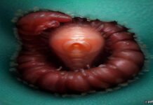





In the world of medical anomalies, few stories capture the public imagination—and trigger a visceral physical reaction—quite like the intersection of parasitology and ophthalmology. Recently, a harrowing case study surfaced involving a 68-year-old patient who sought medical intervention for persistent ocular discomfort, only to discover that his eyes had become a breeding ground for parasitic worms. This incident, while rare, serves as a stark reminder of the complexities of zoonotic diseases and the delicate nature of the human ocular system.
The Clinical Presentation of Ocular Myiasis
The patient, a resident of a rural region, initially presented to an emergency clinic complaining of intense itching, a sensation of a foreign body, and localized inflammation in both eyes. For several days, the discomfort had escalated, leading to excessive tearing and blurred vision. While the medical team initially suspected common conditions such as allergic conjunctivitis or a corneal abrasion, the physical examination revealed something far more unsettling: the presence of multiple, translucent, thread-like organisms moving across the conjunctiva.
Upon closer inspection using a slit-lamp biomicroscope, ophthalmologists identified the intruders as larvae belonging to a species of fly, a condition clinically referred to as ocular myiasis. The worms were not merely present; they were actively migrating across the ocular surface. The urgency of the situation became immediately apparent when the medical team observed that the larvae were in an advanced stage of development. In many cases of parasitic infestation, the window between initial colonization and the hatching or bursting of larvae is narrow, necessitating immediate surgical intervention to prevent permanent damage to the cornea and surrounding tissues.
The Mechanics of an Ocular Infestation
How exactly do these organisms find their way into the human eye? Myiasis is a condition caused by the larvae of various fly species that feed on the host’s necrotic or living tissue. While most common in livestock, human cases can occur, particularly in individuals who spend significant time outdoors in environments where these insects are prevalent. The fly deposits eggs, often through incidental contact near the eye, or via mechanical transmission when a person wipes their eye after touching an infested animal or contaminated surface.
Once the eggs hatch, the larvae thrive in the warm, moist environment of the conjunctival sac. They feed on the secretions and superficial layers of the eye, causing significant mechanical irritation and chemical inflammation. The patient in this case was fortunate; the medical staff acted with extreme precision to remove the larvae one by one using specialized forceps. Each extraction was performed under local anesthesia to ensure the patient remained still, as any sudden movement could have resulted in the larvae burrowing deeper into the ocular structure or causing a rupture of the delicate conjunctival membrane.
The Surgical Challenge and Recovery
The removal process was described by the attending surgeons as a delicate balancing act. Because the larvae were near the point of pupation, their structural integrity was compromised, making them prone to fragmentation if handled incorrectly. If a larva were to rupture during extraction, it could release toxins or bacteria into the eye, potentially leading to severe secondary infections like endophthalmitis—a condition that can lead to permanent blindness if not managed aggressively.
Following the removal of the parasites, the patient underwent a rigorous course of topical antibiotics and anti-inflammatory eye drops to manage the residual trauma. The ocular surface is highly sensitive, and the micro-abrasions caused by the larvae’s hook-like mouthparts required careful monitoring to ensure that secondary corneal ulcers did not develop. Fortunately, the follow-up examinations indicated that the patient’s vision was preserved, and the inflammation subsided without long-term complications. This successful outcome underscores the importance of prompt medical evaluation whenever an unusual sensation occurs in the eye.
Implications for Public Health and Awareness
While stories involving parasitic worms in the eyes are often sensationalized in media, they highlight significant public health themes. For those living in endemic areas, basic hygiene practices—such as washing hands after handling livestock and avoiding direct contact with flies—remain the primary defense against such infestations. Furthermore, the case highlights the necessity for primary care physicians to maintain a high index of suspicion when dealing with patients who present with “foreign body sensations” that do not resolve with standard ophthalmic treatments.
The medical community continues to document these cases to better understand the behavior of these parasites and to refine surgical protocols. As climate change shifts the habitats of various insect species, experts suggest that we may see an increase in the frequency of zoonotic interactions. Consequently, the ability to rapidly diagnose and treat ocular myiasis is becoming an increasingly relevant skill for ophthalmologists worldwide.
Future Outlook
Looking ahead, the integration of advanced imaging technology in clinical settings will likely improve the early detection of ocular parasites, allowing for non-invasive treatment plans before manual extraction becomes necessary. Research into specialized antiparasitic eye drops that can neutralize larvae without damaging human tissue is also ongoing. While the thought of ocular parasites remains deeply unsettling, the advancements in diagnostic precision and surgical techniques ensure that even in the most extreme cases, patients have a high probability of retaining their sight and returning to full health. Education and awareness remain the most effective tools in preventing these rare but distressing medical encounters.
Original reporting: source.